![]()
![]()
Home
![]()
Editorial
![]()
Free
Medical Advice
![]()
Patient Education
![]()
Review
![]()
Interview
![]()
Horizons
![]()
Sections
![]()
News
![]()
Events
![]()
Directory
![]()
Jokes
![]()
Links

The Heart
About Dr Abdul Hafeez Chaudhry
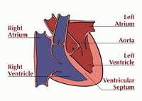
Structure of the Heart
The heart is a muscular organ located just to the left of the breast bone (sternum).
It is about the size of your fist, and this amazing muscle pumps 4300 gallons of blood a
day. The heart has four chambers:
Atria. The top two chambers that receive blood from the body or lungs.
Ventricles. The bottom two chambers. The right ventricle pumps blood to the
lungs to pick up oxygen, The left ventricle pumps blood to the rest of the body and is the
strongest chamber.
Valves. There are four valves in the heart that help to direct blood flow. As
they open and close, the valves produce sounds that can be heard with a stethoscope. The
heart sounds can often tell your doctor about your hearts function.
Function of the Heart
Every cell in your body needs oxygen in order to live and function. The role of the heart
is to deliver the oxygen-rich blood to every cell in the body.The arteries are the
passageways through which the blood is delivered. The largest artery is the aorta, which
branches off the heart and then divides into many smaller arteries. The veins carry the
deoxygenated blood back to the lungs to pick up more oxygen, and then back to the heart
once again. Blood flows continuously through the circulatory system, and the heart muscle
is the pump which makes it all possible!
Coronary Arteries
Your heart, just like all other muscles in the body, needs its own supply of oxygen in
order to function properly. Although its chambers contain blood, the heart receives no
nourishment from the blood inside the chambers. The heart gets its blood supply from the
coronary arteries. The two major coronary arteries (the right coronary artery and the left
main coronary artery) branch off the aorta, and then divide into many smaller arteries
that lie in the heart muscle and feed the heart.
The Matter Of The Heart
The heart you see drawn on the average Valentine is only a rough representation of the
actual structure of the heart. Your heart is actually shaped more like an upside-down
pear. The human heart is primarily a shell. There are four cavities, or open spaces,
inside the heart that fill with blood. Two of these cavities are called atria. The other
two are called ventricles. The two atria form the curved top of the heart. The ventricles
meet at the bottom of the heart to form a pointed base which points toward the left side
of your chest. The left ventricle contracts most forcefully, so you can best feel your
heart pumping on the left side of your chest.
The left side of the heart houses one atrium and one ventricle. The right side of the
heart houses the others. A wall, called the septum, separates the right and left sides of
the heart. A valve connects each atrium to the ventricle below it. The mitral valve
connects the left atrium with the left ventricle. The tricuspid valve connects the right
atrium with the right ventricle.
The top of the heart connects to a few large blood vessels. The
largest of these is the aorta, or main artery, which carries nutrient-rich blood away from
the heart. Another important vessel is the pulmonary artery which connects the heart with
the lungs as part of the pulmonary circulation system. The two largest veins that carry
blood into the heart are the superior vena cava and the inferior vena cava. They are
called "vena cava" because they are the "heart's veins." The superior
is located near the top of the heart. The inferior is located beneath the superior.
The heart's structure makes it an efficient, never-ceasing pump. From the moment of
development through the moment of death, the heart pumps. The heart, therefore, has to be
strong. The average heart's muscle, called cardiac muscle, contracts and relaxes about 70
to 80 times per minute without you ever having to think about it. As the cardiac muscle
contracts it pushes blood through the chambers and into the vessels. Nerves connected to
the heart regulate the speed with which the muscle contracts. When you run, your heart
pumps more quickly. When you sleep, your heart pumps more slowly.
Considering how much work it has to do, the heart is surprisingly small. The average adult
heart is about the size of a clenched fist and weighs about 11 ounces (310 grams). Located
in the middle of the chest behind the breastbone, between the lungs, the heart rests in a
moistened chamber called the pericardial cavity which is surrounded by the ribcage. The
diaphragm, a tough layer of muscle, lies below. As a result, the heart is well protected.
To monitor the heart, scientists can use x-ray or scanning technology to get a picture. To
really explore the heart, scientists have to perform surgery. Heart surgery is very risky
because the heart's pumping action is so critical for survival. If the heart stops
pumping, the body cannot survive. Before beginning heart surgery, doctors connect the
patient to a machine that pumps the blood for the heart. Only then is it safe for the
doctor to stop the heart in order to operate.
Courtesy,
Franklin Institute online