![]()
Home
![]()
Editorial
![]()
Free
Medical Advice
![]()
Patient Education
![]()
Review
![]()
Interview
![]()
Horizons
![]()
Sections
![]()
News
![]()
Events
![]()
Directory
![]()
Jokes
![]()
Links
Traumatology
Important definitions
1- Trauma: Diseased condition of the body produced by external
violence. It includes emotional and mental
stress resulting into a disorder.
2- Wound: It is not legally defined in Pakistan.
Medically, it is defined as breech in the integrity of a tissue caused by force/energy.
3- Injury: (legal term) Defined in Sec.44 PPC.
" Injury denotes any harm whatsoever caused illegally to a person in body, mind,
reputation and property ".
4- Hurt: (legal term) Defined in Sec 332 PPC. "
Causing of pain, harm, disease, infirmity, injury or impairing, disabling, dismembering
any organ of the body or part there of without causing death ".
Classification of Wounds
Any doctor may be asked to examine a person who has been wounded, particularly in
casualty.
Forensic physicians and pathologists are also asked to examine wounds, whether they are serious or trivial, and whether the injured person is alive or dead.
The identification and description of wounds may have serious medico-legal implications at a later stage, and often after some considerable time has passed since the wounding. It is therefore essential that different types of wounds can be correctly identified and described, with a full description being made in notes taken at the time of, or shortly after the examination ('contemporaneous notes').
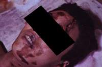
A wound is the term given to tissue damage caused by mechanical force. This includes wounds caused by stabbing, blunt trauma (punching, kicking, beating etc), strangling, biting, shooting, falling from a height, being hit by a vehicle, and blast trauma from explosives.
Descriptions of wounds must include, the nature of the wound, ie whether it is a bruise, abrasion or laceration etc the wound dimensions, eg length, width, depth etc. It is helpful to take a photograph of the wound with an indication of dimension (eg a tape measure placed next to the wound), and for measurements to be taken of the wound as it appears first, and then with wound edges drawn together (if it is a laceration etc). the position of the wound in relation to fixed anatomical landmarks, eg distance from the midline, below the clavicle etc the height of the wound from the heel (ie ground level) - this is particularly important in cases where pedestrians have been struck by motor vehicles
The following is main types of wounds:
bruises/ contusions
abrasions
lacerations
incised wounds
stab wounds
defence injuries
1- Bruises/ Contusions
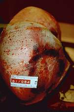
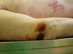
Bruises are caused by blunt trauma / injury to tissues, resulting in damage to
blood vessels beneath the surface. Blood leaks out ('extravasation') into surrounding
tissues from damaged capillaries, venules and arterioles. Bruises may be surface bruises,
or deeper within tissues or organs.
Unlike abrasions, the characteristics of the object causing a bruise cannot easily be
determined, because blood tends to spread out in a diffuse manner from the site of injury,
particularly along fascial planes. Indeed, this phenomenon results in the apparent
'shifting' of bruises after time. For example, a scalp injury may result in a black eye.
Bruses may also 'appear' after some days due again to the same phenomenon of blood
tracking along tissue planes, and pathologists often re-examine a body again tolook for
such bruising.

Intra-dermal bruises, however, provide an exception to this general rule, as they are
superficial - lying just under the epidermis. In this case, there may be good correlation
between the bruise seen and the characteristics of the causative object.
Additional terms are used for bruises of a particular size,
ecchymoses - smaller than a few milimeters
petechiae - pinpoint bruises. (However, this type of bruise or haemorrhage are usually due
to venous engorgement, for example in asphyxia, or in defects in blood coagulation such as
Disseminated Intravascular Coagulation (DIC) than trauma). Light trauma may produce
petechial haemorrhage, but those caused by venous congestion etc are more diffuse in
nature.
A blow from an object may give rise to a combination of injuries, such as a bruise with an abrasion etc, and different parts of the body are more susceptible to bruising than others. For example, the skin over the eyelids bruises easily, whilst the tougher palmar surface or plantar surface rarely bruises.
'Tramline' bruises consist of two parallel linear bruises separated by a paler, undamaged section of skin. This type of injury occurs when the skin is struck with a rod shaped object, which sqeezes blood from the vessels at the point of inpact, thus emptying them and preventing them from leaking blood. The edges of the wound are stretched, and blood vessels are torn, causing blood to leak into the surrounding tissues. A similar phenomenon is seen when the injury is caused by a hard spherical object, such as a squash ball !
The positioning of bruising is often significant, in that a multiple row of spherical/ disc shaped bruises may be seen when an attempt is made to strangle someone with bare hands. The bruises are caused by the attacker's fingertips pressing into the skin.
Bruises change colour over time, because of the degradation of haemoglobin in the blood. However, the timescale of this degradation is not fixed, and it is therefore possible only to give a rough estimation of the age of the bruise. Colour changes are,
dark blue/ purple (fresh)
blue
brown
green
yellow
In general, small bruises on an otherwise fit and healthy person, could pass through the spectrum of colour changes between 72 hours and 1 week. The more extensive, or deep seated the bruise, the longer it will take to dissapear. If a bruise is brown/ green or yellow it is likely that the injury is at least 18 hours old.
An important observation is the colour of multiple bruises on the same person - markedly different coloured bruises suggest that they have been caused at different times, and may indicate signs of chronic abuse, such as of an infant etc.
A possible line of defence for a person charged with homicide could be that the bruises were not in fact made during life, but are post-mortem artifacts. However, it is very difficult to produce a bruise in a dead person, because it is very much a 'vital reaction' to injury. It may be possible to produce a bruise following very severe trauma in an area of post-mortem lividity (where blood has drained to dependant parts of the body under the influence of gravity), and if there is any doubt the pathologist should examine the 'bruise' histologically. However, in the absence of an organising haematoma, histology may only
show thepresence of haemosiderin, and even then only after approximately 48 hours.
In forensic autopsies, it is important that all possible signs of trauma are specifically looked for and excluded. This includes a thorough search for bruising, including that which is beneath the skin surface. Forensic pathologists use specialist dissection
techniques to expose deeper structures, and this may entail removing all skin off whatever structure is being looked at.
2- Abrasions
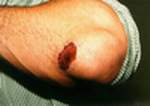
An abrasion is a superficial injury, commonly known as a 'graze' or 'scratch'.
This type of wound damages only the epidermis (uppermost skin layer), and should not therefore bleed. However, abrasions do usually extend into the dermis causing slight bleeding.
Abrasions are commonly caused by a 'glancing' impact across the surface of the skin, but if the force is directed vertically down onto the skin surface it may be termed a 'crush' injury.


These wounds are seen where an object has struck the skin (eg a blow from a fist), or
where the injured person has fallen onto a rough surface, such as road.
Abrasions may be 'linear', or commonly known as a single 'scratch', whereas if a broader surface is affected, it is called a 'graze' or 'brush abrasion' (eg where a motorcyclist is thrown from their vehicle, and comes into contact with the road surface in a skidding fashion). Such an abrasion often covers a relatively large area of skin, and is often called a 'friction burn' in lay language.
If the surface of an abrasion is examined closely, for example with a
hand-held magnifying glass, the direction of force can often be determined, from the torn
epidermis. Strands are drawn towards the end of the injury, and are 'heaped up'. The edges
of the wound may also be ragged and directed towards the end of the wound.

Of particular importance in the forensic setting is the fact that abrasions can retain
much of the surface characteristics of the object that caused the wound. For example,
there may be a patterned abrasion caused by an element of a vehicle involved in a
'hit-and-run' (such as that made by a radiator grill or bumper), and if the abrasion has
been fully documented and photographed (with a scale) and the suspected vehicle is
subsequently recovered, the two may be matched up.
(Lacerations & Incised Wounds) Click here.
